Magnetic resonance imaging (MRI) is now widely applied in clinical diagnostics. Its advantages—non-radiative safety, flexible tomographic scanning from any angle, and the ability to capture proton density, relaxation, weighted images, and multiparametric features—make it one of the most powerful diagnostic tools today. However, in clinical practice, relaxation times of different tissues or tumour tissues sometimes overlap, complicating diagnosis. To address this, contrast agents were developed to enhance signal contrast and improve resolution. By altering local tissue relaxation properties after injection, contrast agents increase image contrast and diagnostic accuracy.
Contrast agents are chemically synthesised substances with higher density than living tissue. They do not generate signals themselves, but by altering the relaxation efficiency of water protons in local tissues, they create contrast with surrounding tissue to achieve visualisation.
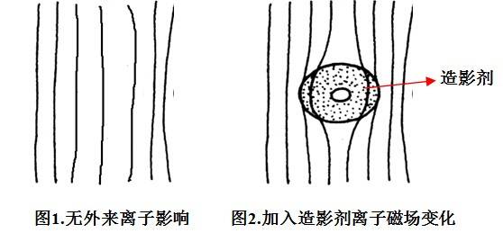
Magnetic substances such as Fe, Mn, and Gd contain multiple unpaired electrons. When near resonating hydrogen atoms, they alter the local magnetic field, markedly shortening T1 and T2 relaxation times. Contrast agents therefore change the relaxation rate of water protons in tissues, increasing contrast and resolution between healthy and diseased sites, providing more diagnostic information (see Figures 1 and 2).
MRI contrast agents are usually paramagnetic or superparamagnetic. They interact magnetically with hydrogen nuclei. Once inside the body, they alter longitudinal relaxation rates (1/T1) and transverse relaxation rates (1/T2). Under the influence of paramagnetic substances, diamagnetic and paramagnetic contributions are additive:
(1/Ti)obs = (1/Ti)dia + (1/Ti)para (where i = 1, 2).
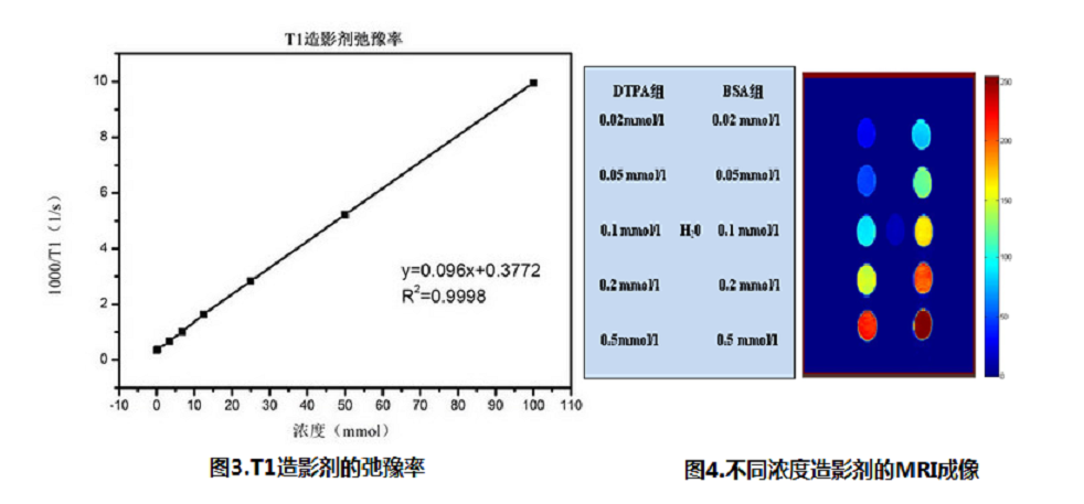
In the absence of solute–solute interactions, the solvent relaxation rate is linearly related to the concentration (mol/L) of added paramagnetic substances:
(1/Ti)obs = (1/Ti)dia + Σri[C] (i = 1, 2).
Here, ri is the relaxivity (mmol/L·s) of the paramagnetic substance, and Σ refers to contributions from all contrast agents in solution. T1-type agents (e.g., Gd-based complexes) brighten regions on imaging, also called positive contrast agents. T2-type agents (e.g., Fe3O4-based superparamagnetic particles) darken regions, known as negative contrast agents.
Relaxivity is one of the most important indicators of MRI contrast agents. High-relaxivity agents achieve the desired effect with minimal dosage. In this field, Niumag has developed compact MRI analysers capable of measuring T1 and T2 relaxation times and imaging tube samples. These systems provide both quantitative and qualitative data, offering researchers fast, reliable tools for the evaluation and optimisation of new contrast agents.
In-vivo evaluation of contrast agents:
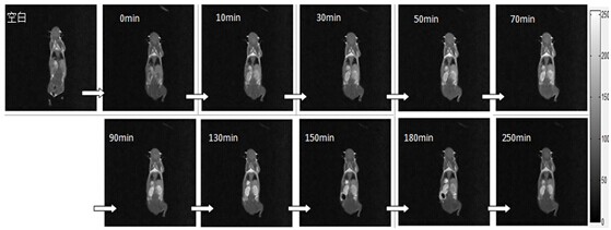
Monitoring metabolism of a contrast agent in the kidney: in mice, renal metabolism lasted over 250 minutes, with peak activity around 130 minutes post-injection.
In biomedical research, low-field NMR can support the following applications:
1) Contrast-agent research: relaxivity, efficacy evaluation, in-vivo metabolism studies.
2) Subcutaneous tumour studies: contrast effects, drug targeting, tumour efficacy assessment.
3) In-situ tumour research: localisation, metastasis assessment, size measurement.
4) Body composition analysis in conscious animals: lean mass, fat mass, free water content, fat distribution.
 Scan QR Code
 Scan QR Code
Phone: 400-060-3233
After-sales: 400-060-3233


Back to Top