To investigate the MRI contrast-enhanced imaging of subcutaneous tumours in mice and study the metabolic process of the contrast agent.
2.1 Experimental Materials
Mouse (#20), MRI contrast agent.
2.2 Experimental Instruments
Small-animal MRI system, magnet strength 0.5 T, mouse coil diameter 40 mm.
2.4 Experimental Method
Using Niumag MRI software, imaging was performed with an MSE sequence. After injection of the MRI contrast agent, T2-weighted imaging was carried out according to the study design.
3.1 Mouse #20 received intravenous tail-vein injection of the MRI contrast agent, followed by observation of the tumour site using T2-weighted imaging.
Post-injection T2-weighted tumour images were obtained as shown below. Results indicated that between 24–72 minutes after injection, the tumour area showed marked darkening. By 120 minutes, tumour brightness had partially recovered.
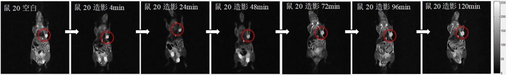
Fig. 3 T2-weighted images of mouse #20 after intravenous injection of contrast agent (grayscale)
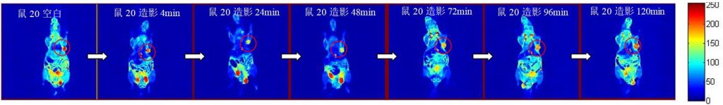
Fig. 4 T2-weighted images of mouse #20 after intravenous injection of contrast agent (pseudo-colour)
 Scan QR Code
 Scan QR Code
Phone: 400-060-3233
After-sales: 400-060-3233


Back to Top