
Bauhinia seeds;
NMI20-015V-I, resonance frequency: probe coil with a 15 mm diameter;
T2 Relaxation Test: Bauhinia seeds were soaked in water for set intervals (0h, 1h, 2h, 4h, 6h, 8h, 12h, 16h, 20h, 24h, 48h);
MRI Test: Proton density imaging was performed on samples of Bauhinia seeds soaked for different durations.
IV. Experimental Parameters
CPMG: SF = 22 MHz, O1 = 107966 Hz;
MRI: SE sequence.
Soaked-seed T2 spectra: CPMG sequence was used to test water-saturated seeds, and the SIRT algorithm was applied to invert and generate T2 spectra;
MRI: Coronary-plane imaging of Bauhinia seeds was acquired using NMR imaging software with an SE sequence;
(1) Analysis of T2 spectra of Bauhinia seeds at different soaking times
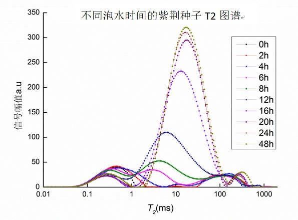
The spectra reveal clear trends across the first three peaks, representing bound water, less-mobile water, and free water. After soaking, notable changes appear: the normalized T2 spectra highlight shifts in peak positions and areas. Area A22 showed the strongest correlation with the total area (Atotal), representing the dominant variation during seed hydration. Linear fitting of Atotal to water absorption rate produced strong results, with seeds reaching hydration equilibrium after about 24 hours.
(2) Seed Imaging Experiments
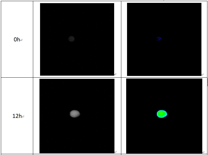
The coronal images clearly show the hydration process from 0h to 12h: water uptake begins at the right-side embryo and gradually spreads throughout the seed before equilibrium is reached. Overall, LF-NMR analysis effectively reveals the distribution of water states in Bauhinia seeds, tracks the migration pathway during soaking, and identifies the time required to reach equilibrium.
 Scan QR Code
 Scan QR Code
Phone: 400-060-3233
After-sales: 400-060-3233


Back to Top