
Walnut, also known as Juglans, is rich in walnut oil, protein, carbohydrates, minerals, and vitamins. Walnut oil contains a high level of unsaturated fatty acids, including palmitoleic acid, oleic acid, linoleic acid, and linolenic acid. These unsaturated fats are beneficial in lowering cholesterol and blood lipids and play a significant role in preventing atherosclerosis and thrombosis.
Due to its higher market value compared to other vegetable oils, some manufacturers intentionally adulterate walnut oil with cheaper, visually similar oils such as soybean oil, corn oil, or sunflower oil to cut costs and maximize profit. This poses a significant challenge to quality assurance. Traditional quality inspection methods are time-consuming, chemical-intensive, and not suitable for real-time monitoring. Moreover, conventional instrumental detection techniques often show poor correlation with standard indicators or yield unsatisfactory results.
Therefore, developing a reliable method for detecting adulterated walnut oil is critical for quality control and market regulation.
Low-field nuclear magnetic resonance (LF-NMR) is increasingly used in the food industry due to its advantages of speed, accuracy, non-destructiveness, and no need for chemical reagents. In this study, LF-NMR is used to analyze pure walnut oil and samples adulterated with soybean, corn, or sunflower oil. Coupled with chemometric methods, CPMG echo data and known adulteration ratios are analyzed to assess walnut oil quality.
Case Study 1: Pure Oil Sample Analysis
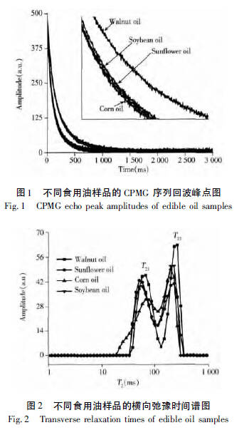
Walnut oil, corn oil, soybean oil, and sunflower oil were tested under identical experimental parameters. The CPMG relaxation decay signals and T2 relaxation results were processed using professional software for T2 inversion. The resulting data were plotted using Origin 8.5 software to generate the CPMG decay curves (Figure 1) and relaxation spectra (Figure 2).
Case Study 2: Adulterated Oil Sample Analysis
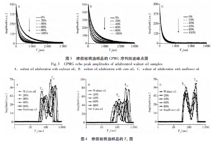
Under the same testing conditions, 99 walnut oil samples adulterated with varying proportions of soybean, corn, and sunflower oils were analyzed using LF-NMR. The resulting CPMG decay curves and T2 relaxation spectra are shown below.
As the level of adulteration increased, the echo signal decayed more rapidly—indicating a higher relaxation decay rate (Figure 3). Additionally, both the T21 and T22 peaks shifted to the left (Figure 4), reflecting changes in molecular mobility and interaction dynamics caused by adulteration.
For principal component analysis and further technical details, please refer to:
“Rapid Detection of Adulterated Walnut Oil Using Low-Field NMR Combined with Chemometric Methods,” Journal of Instrumental Analysis, Vol. 34, No. 7, July 2015.
 Scan QR Code
 Scan QR Code
Phone: 400-060-3233
After-sales: 400-060-3233


Back to Top